Monday, December 31, 2012

Experiences of caring for patients with lymphedema and dementia

Experiences of caring for patients with lymphedema and dementia

Dec 2012

[Article in Japanese]

Source

The Palliative Care Units, Sanshu Hospital.

Abstract


Recently, the importance of prevention and care of lymphedema patients has been recognized. To improve edema reduction and skin condition, continued care is necessary. However, everyday care requires time and effort, and patients may neglect the required routine. Therefore, it is necessary for patients to recognize the importance of lymphedema and acquire the knowledge and skills to care for their condition. In the case of patients with dementia, it is important to ensure 1) comfortable care for the patients, 2) flexibility of care according to the physical and mental state of the patients, and 3) education of visiting home care staff.
**As more and more lymphers are aging, caring for those of us who have/get dementia will be a significant challenge.**

Angiosarcoma in a patient with congenital nonhereditary lymphedema.


Angiosarcoma in a patient with congenital nonhereditary lymphedema.


Nov 2012

Source

Divison of Anatomic Pathology, Mayo Clinic, Rochester, Minnesota 55905, USA.

Abstract


Angiosarcoma is an uncommon but aggressive tumor of endothelial origin that may occur in the upper extremities of patients with postmastectomy lymphedema (Stewart-Treves syndrome) as well as in other regions. We present an unusual case of angiosarcoma associated with congenital nonhereditary lymphedema in an 18-year-old man. Our case underscores the need for a careful clinical examination and shows the importance of appropriate sampling and thorough pathologic examination of suspicious areas to exclude the presence of a malignant process.


For further information:


Wednesday, December 26, 2012

Control of lymphorrhea and treatment of warty excrescences in elephantiasis.


Control of lymphorrhea and treatment of warty excrescences in elephantiasis.


2012

Source

Cardiology and Cardiovascular Surgery Department, Faculty of Medicine School of São José do Rio Preto (FAMERP), Avenida Brigadeiro Faria Lima, 5416 Vila São Pedro, 15090-000 São José do Rio Preto, SP, Brazil ; Vascular Laser Center, Clínica Godoy, 1306 Avenida Constituição, 15025-120 São Jose do Rio Preto, SP, Brazil.

Abstract


The aim of this study is to report the control of lymphorrhea in the intensive treatment of elephantiasis, using an Unna boot. The case of a 29-year-old female patient is reported. This young patient evolved with the more serious form of lymphedema, elephantiasis, after surgical treatment of an abdominal neoplasm and radiotherapy. Warty excrescences were present on both legs and genitalia where lymphorrhea was constant. The patient arrived at the Godoy's Clinic for treatment. She was weighed and perimetric evaluations were made at the start of treatment and thereafter every day during an intensive outpatient treatment of eight hours daily for three weeks. Treatment included manual lymph drainage, mechanical lymph drainage using the RA Godoy device, and the continuous use of compression stockings with adjustments made every three hours. An Unna boot was employed as compression at sites of dermal lesions (warty excrescences) with overlapping use of individualized compression stockings that were individually adapted. The Unna boot was renewed every two days during the first week and every 3 days during the second and third weeks. By the end of this course of treatment, most of the warty excrescences had reduced in size or even disappeared and the lymphorrhea was controlled. 

Full Text Article with images:

Tuesday, December 25, 2012

Management of unusual genital lymphedema complication after Fournier's gangrene: a case report.


Management of unusual genital lymphedema complication after Fournier's gangrene: a case report.


**Editor's note:  I am posting this as I just can't believe they did this. Perhaps it was

necessary due to the extent of the gangrene tissue damage. Otherwise, remember
- those of you who are men here - there are other surgical options much less
brutal and intrusive.  Pat**                 


Management of unusual genital lymphedema complication after Fournier's gangrene: a case report.


Dec 2012

Abstract


BACKGROUND: Fournier's gangrene is a bacterial infection characterized by necrotizing fasciitis, skin and soft tissue involvement, and eventually myositis of the perineal region. Aggressive debridement of devitalized tissue and overlying skin is of paramount importance, but often leaves large defects to be reconstructed. The present case reports successful extensive perineal defects coverage following Fournier's gangrene and management of subsequent penile lymphoedema impairing sexual function in a young patient.

CASE PRESENTATION:

Following perianal abscess drainage, a healthy young man presented with scrotal pain. Fournier's gangrene was diagnosed and treated with multiple surgical debridements. Tissue excision extended through the entire perineal area, base of the penile shaft, lower abdominal region, the inner thighs, and gluteal region, corresponding to 12 % of the total body surface area. After serial debridements and negative pressure dressings, the defect was covered by two stages of skin grafting. Graft take was 90 %. Healing was achieved without hypertrophic or retractile scar. However, chronic penile lymphedema remained and was first treated with compressive garments for 2 years. Upon failure of this conservative approach, we performed a circumcision, but only a "penile lift" allowed a satisfactory esthetical and functional result.

CONCLUSION:

Fournier's gangrene can be complicated by a chronic lymphedema of the penis. Conservative treatment is likely to fail in severe cases and can be treated surgically by "penile lift".

Saturday, December 22, 2012

Complex Decongestive Physiotherapy Treats Skin Changes like Hyperkeratosis Caused by Lymphedema.


Complex Decongestive Physiotherapy Treats Skin Changes like Hyperkeratosis Caused by Lymphedema.


2012

Source

School of Physical Therapy and Rehabilitation, Abant Izzet Baysal University, 14280 Bolu, Turkey.

Abstract


Lymphedema is a chronic, progressive, and often debilitating condition. Primary lymphedema is a lymphatic malformation developing during the later stage of lymph angiogenesis. Secondary lymphedema is the result of obstruction or disruption of the lymphatic system, which can occur as a consequence of tumors, surgery, trauma, infection, inflammation, and radiation therapy. Here, we report a 64-year-old woman presenting with hyperkeratosis, a lymphedema due to metastatic uterus carcinoma. In this paper, we present the effects of complex decongestive physiotherapy on lymphedema and hyperkeratosis. For further information:

Wednesday, December 19, 2012

Blood test accurately detects lymphedema, study shows


Blood test accurately detects lymphedema, study shows


Dec 18, 2012

Scientists at the Stanford University School of Medicine have identified a set of proteins circulating in blood whose levels accurately flag the presence of lymphedema. The findings, to be reported Dec. 18 in PLoS ONE, spur optimism that this common but relatively neglected condition, which affects an estimated 10 million people in the United States, finally will be amenable to detection (and, eventually, treatment) with 21st-century techniques.

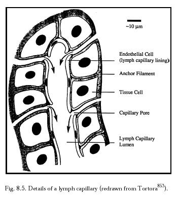
Lymphedema is an often-painful inflammatory condition resulting from the blockage of lymphatic vessels that ordinarily drain fluid from the tissues throughout the body. In the developed world, lymphedema most often arises as an unintended consequence of radiation therapy for cancer. For example, about one in four breast-cancer survivors eventually develops lymphedema, said Stanley Rockson, MD, professor of cardiovascular medicine and the study's senior author. Numerous other factors, including parasitic infections endemic in some developing countries, can cause it as well, he said. 

The blunting of normal immune-cell flow due to lymphatic-vessel blockage helps to trigger the buildup of fluid within the affected area of the body, along with thickening of the skin, profound inflammation, accumulation of fibrous tissue, excessive blood-vessel formation and a marked expansion of the fatty layer beneath the skin. 

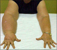
By the time the main symptom—swelling of one or more limbs—is detectable, the condition may have gotten such a foothold that it becomes difficult or impossible to reverse, at least given current treatment options, Rockson said. 

The only known way to diagnose lymphedema now is via physical inspection, and all too often it is misdiagnosed or overlooked altogether. But the biological events underpinning this condition may be present five years or more before symptoms become evident, said Rockson. Moreover, there are no effective drugs for combating lymphedema, just costly, time-consuming and annoying physical therapy, which virtually never completely eliminates the symptoms. While physical therapy can arrest progression and reduce swelling by as much as half, the condition typically remains a long-term problem. "Lymphedema virtually never just goes away on its own," said Rockson. Indeed, it tends to progress in severity over time, whether it is treated or not.

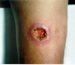
The irreversible skin thickening, joint immobility, scarring, increased susceptibility to infection and other consequences of chronic lymphedema can leave patients discomfited for life, all too often resulting in social withdrawal, body-image deterioration and other quality-of-life issues. "This is especially ironic in the case of cancer survivors who have endured difficult life-saving interventions, only to find that now cured, they're unable to enjoy their lives," said Rockson, who is the Allan and Tina Neill Professor of Lymphatic Research and Medicine and chief of consultative cardiology at the medical school. 

For this study, Rockson and his associates obtained skin-biopsy samples from both lymphedematous and normal tissue of 27 patients. Using advanced molecular methods, they compared each patient's diseased tissue with that same patient's healthy tissue to see which genes—the recipes for the myriad proteins produced in our bodies—were more actively engaged in the generation of their respective protein products in diseased versus healthy tissue. Thousands of genes fit the bill. Then the investigators narrowed their search to the overproduced proteins themselves, in particular ones that were already known to circulate throughout the bloodstream of all people, including healthy ones, and for which fast, commercial blood tests already exist. 

Statistical modeling indicated a panel of tests that measured six separate proteins' levels in study subjects' blood was able to distinguish the lymphedematous patients from control subjects who did not have lymphedema. None of these six proteins was predictive by itself. But in aggregate, their presence at certain levels and ratios appeared to serve as a biological fingerprint, or biomarker, for lymphedema. 

Interestingly, all six proteins are well-known, and each is associated with one or another of chronic lymphedema's hallmark biological features: accumulation of fibrous deposits, stimulation of fat-cell activity, inflammation and lymphatic-vessel formation and repair. "These biomarkers may themselves lead us to valuable pharmaceutical targets," said Rockson. 

To determine their six-protein biomarker-panel's validity, Rockson's group collected blood from a new cohort of 36 lymphedematous and 15 healthy adults, extracted blood samples and tested them with the panel. The test distinguished those with lymphedema from healthy subjects with an accuracy approaching 90 percent—good enough for use as a clinical 
diagnostic tool and a vast improvement over current detection methods, said Rockson. 

"This is a significant development," he said. 

Because levels of the six proteins begin to climb early in the course of the disease, such a test should be valuable in determining risk for, or the onset of, lymphedema long before symptoms occur—which in turn would mean earlier, appropriate therapeutic intervention, perhaps in time to spare patients from the condition's most-damaging effects or even reverse its course. "In addition," Rockson said, "a standardized, accurate bioassay for lymphedema could help to pave the road for future human clinical trials of drugs to treat it." Monitoring trial subjects at the molecular level with a lymphedema-detecting blood test could provide early evidence regarding whether an experimental treatment is working. Rockson is involved in conducting clinical trials of pharmaceutical agents for lymphedema, and hopes to use the new test in those trials. Journal reference: PLoS ONE Provided by Stanford University Medical Center 

Medical express

The published study itself:

PLoS One

Monday, December 17, 2012

Lymph Glands or Lymph Nodes


Steady Health


Anatomy for Nurses

Physiotherapeutic stimulation: Early prevention of lymphedema following axillary lymph node dissection for breast cancer treatment

Physiotherapeutic stimulation: Early prevention of lymphedema following axillary lymph node dissection for breast cancer treatment

Authors: Almir J. Sarri, Sonia M. Moriguchi, Rogério Dias, Stela V. Peres, Eduardo T. Da Silva, Kátia H. Koga, Ângelo G.Z. Matthes, Marcelo J. Dos Santos, Euclides T. Da Rocha, Raphael L. Haikel

Affiliations: Department of Physiotherapy, Barretos Cancer Hospital, São Paulo, Brazil. almirsarri@hotmail.com

Doi: 10.3892/etm_00000024

Pages: 147-152

Abstract:

The aim of this study was to confirm the effectiveness of early physiotherapeutic stimulation for lymphatic flow progression in patients with breast cancer undergoing axillary dissection. This was a randomized experimental study on 22 patients who underwent lymphoscintigraphy in their arms on two different occasions, firstly without stimulation and secondly after randomization into two groups: without physiotherapeutic stimulation (WOPS; n=10) and with physiotherapeutic stimulation (WPS; n=12). The lymphoscintigraphy scan was performed with 99mTc-phytate administered into the second interdigital space of the hand, ipsilaterally to the dissected axilla, in three phases: dynamic, static, and delayed whole body imaging. Physiotherapeutic stimulation was carried out using Földi's technique. In both groups, images from the two examinations of each patient were compared. Flow progression was considered positive when, on the second examination, the radiopharmaceutical reached areas more distant from the injection site. Statistical analysis was used to evaluate frequencies, percentages and central trend measurements, and non-parametric tests were conducted. Descriptive analysis showed that the WPS and WOPS groups were similar in terms of mean age, weight, height, body mass index and number of lymph nodes removed. There were statistically significant associations between physiotherapeutic stimulation and radiopharmaceutical progression at all three phases of the study.

Early physiotherapeutic stimulation in breast cancer patients undergoing radical axillary dissection is effective, and can therefore be indicated as a preventive measure against lymphedema. 

Lymphnode Stimulation Front and Back Illustrated

Lymphnode Stimulation Front and Back Illustrated


Sunday, December 16, 2012

Lymphatic Drainage

Lymphatic Drainage


Optimum Health Clinic


Lymphatic Drainage Legs


Lymphatic Drainage Arms


Lymphatic Drainage Head


Junior Dentist

Lymphatic Drainage Head and neck


Darmouth EDU


Lymphatic Drainage neck


Mosby's Dental Dictionary/Answers


Lymphatic Drainage Map

Lymphatic Capillaries

Lymphatic Capillaries


Interstitial Fluid


Nanomedicine


Description:
Lymph capillaries are tiny thin-walled vessels that are located in the spaces between cells throughout the body. They collect excess fluid from cells and recycle it for use in the circulatory system.


Clipart.uk

Compare the blood capillary below to the above lymph capillary

Valves in Lymph Vessel

Valves in Lymph Vessel

right lymphatic duct with internal valve

Tutor Vista


Xenpod

Lymphatic vessel

Lymphedema Webs.com


Compare the lymphatic vessel to the Blood vessel

Blood Vessel Diagram

Sunday, December 09, 2012

Lymph Node anatomy

Lymph Node anatomy

Lymph Node Structure

Lymph Node Structure

Lymph Node Clusters

Upper Lymph Nodes


Lymph Node Regions


Real Complications of Lymphedema


Real People Real Complications of Lymphedema
Perhaps after Barbara’s article,  it is time to post some illustrations of what really does happen to those of us with lymphedema.
Think this doesn’t apply to you?
Remember if you have ever had any of these experiences you are at risk and a possible victim of lymphedema: 
1.) Lymph node removal for biopsies
2.) Serious infections that include lymphangitis, cellulitis or erysipelas.
3.) Deep invasive wounds that might tear, cut or damage the lymphatics.   
4.) Radiation treatments, especially ones that are focused in areas that might contain “clusters” of lymph nodes
5.) Morbid obesity can cause secondary lymphedema by “crushing” the lymphatics
6.) Serious burns, even intense sunburn 
7.) Infection of the microscopic parasite filarial larvae, though this is more common in tropical countries
8.) For primary lymphedema any person who has a family history of unknown swelling of a limb
9.) Spider or insect bites
10.) Surgeries that cut or damage the lymph system.
COMPLICATIONS
 1.  Infections such as cellulitis, lymphangitis, erysipelas. This is due not only to the large accumulation of fluid, but it is well documented that lymphodemous limbs are localized immunodeficient.                       
2.  Draining wounds that leak lymphorrea which is very caustic to surrounding skin tissue and acts as a port of entry for infections.
          
3.  Increased pain as a result of the compression of nerves usually caused by the development of fibrosis and increased build up of fluids.
4.  Loss of Function due to the swelling and limb changes.
         
          
5.  Depression - Psychological coping as a result of the disfigurement and debilitating effect of lymphedema.
6.  Deep venous thrombosis again as a result of the pressure of the swelling and fibrosis against the vascular system. Also, can happen as a result of cellulitis, lymphangitis and infections.
7.  Sepsis, Gangrene are possibilities as a result of the infections.
      
8.  Possible amputation of the limb.
 
9.  Pleural effusions may result if the lymphatics in the abdomen or chest are to overwhelmed to clear the lung cavity of fluids. I just had surgery for this as my lungs continue now to fill up every three weeks. Hopefully, the surgery can prevent it.        
      
10. Skin complications such as splitting, plaques,susceptibility to fungus and bacterial infections.
                                              
Elephantiasis nostras verrucosa     Lymphomatoid Papulosis        Hyperkeratosis, Papillomatosis
11. Chronic localized inflammations.


12. Angiosarcoma, a cancer of the soft tissues
     
Cancers – New research is indicating that those of us with lonh standing lymphedema run a 1 in 10 risk factor for these cancers
13. Lymphangiosarcoma which is a rapidly progressive, non curable cancer of long term lymphedema  patients.  Almost always fatal within six months of diagnosis
14. Lymphoma, new research indicates a possibility of this with hereditary lymphedema.  I have been diagnosed with two forms of lymphoma.     
                                                                                                               Skin lymphoma
15. Kaposi’s Sarcoma, another cancer that can and does arise from lymphedema.
16. Septic arthritis  - type of arthritis triggered/caused by systemic infections, cancers and more
                        
Septic arthritis in knee                Septic arthritis in hand